| |
|
|
 |
|
|
|
||||||
| ||||||
Microscopy, in its many forms, is one of the most important techniques for the analysis of small objects. During the last few decades this technique has expanded in several directions. Currently these include the disciplines known as Scanning Tunnelling Microscopy, Electron Microscopy, Atomic Force Microscopy, Soft X-Ray Microscopy, Laser Scanning Confocal Microscopy and Optical Microscopy. Each of these techniques has certain advantages over it competitors, but only one, Optical Microscopy, has been in the game since the very beginning.
Optical Microscopy cannot compete with the previously mentioned techniques in resolution, but it does have several advantages that explain its ever present popularity. The technique is relatively inexpensive to set up and equally easy to use. Its reliability is unmatched and requires little or no sample preparation. In addition, performance is possible at room temperature and atmospheric pressure allowing a much wider range of possible applications. Of coarse all these advantages come with a price, and this is the lack of resolution. Due to the diffraction limit placed on waves of light in free space, the resolution obtainable is no less than about 300nm. It is precisely this reason that forced microscopy to be performed at shorter wavelengths. This shortfall of optical microscopy lead to the invention of the Electron Microscope
Scanning Near-field Optical Microscopy is a technique that allows us to achieve resolution an order of magnitude below normal Optical Microscopy by "cheating" the diffraction limit. Since it is essentially Optical Microscopy it retains many of the advantages while simultaneously eliminating the major disadvantage. The resolution of this system is governed primarily by the size of the aperture used to collect the light. Hence by fabricating apertures smaller than the wavelength of light used, we obtain sub-wavelength resolution.
 This has uses in atom
optics with its predicted ability to focus an atomic
beam. There is a phase singularity in the focal region of a
lens that has only been observed with far field interference.
This has uses in atom
optics with its predicted ability to focus an atomic
beam. There is a phase singularity in the focal region of a
lens that has only been observed with far field interference.
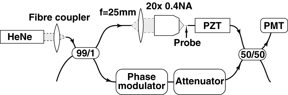
It is also possible to produce interference by mixing light from two optical fibres. This can be done with optical couplers which let the evanescent fields surrounding one optical fibre enter a second fibre which is held in close proximity. This means that light from one fibre is mixed between two. When both fibres have light coming into them, what you get out the end is the interference between the two. The optical fibre interferometer uses one coupler to split a light wave travelling down one fibre into two, and a second coupler to mix these two back together.
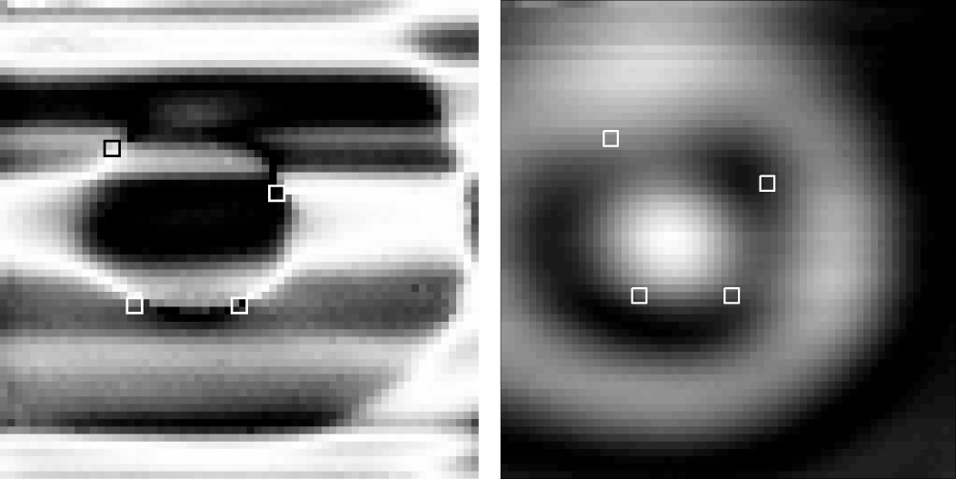
Here is a measurement of the phase structure near the focus of a high
numerical aperture lens. From left to right: cosine of phase,
intensity, greyscale of phase around phase singularity vortex at
bottom left, and shaded surface of same. Left two images are 12x12 microns.

| | Return to Optics Group home page
|
| | Return to School of Physics home page |
![[School of Physics - Optics Group]](../its1.gif)